Horizon Health offers complete radiology services with a commitment to
excellence and patient-centered care.
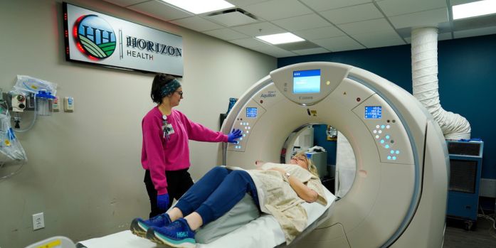
Our next-generation technology exceeds industry standards in all services
offered. We provide faster scan times, quieter experiences, non-contrast
imaging, and same-day test results with an emphasis on patient comfort
and satisfaction.
Equipment modernization includes artificial intelligence for higher quality imaging.
An MRI can be performed on patients with MRI-conditional or MRI-safe implants,
including certain pacemakers, defibrillators, and spinal cord stimulators,
after following strict screening and safety protocols.
One of the widest bores in the industry
Quietest magnet on the market
MRI theater, stereo sound
Comfort lighting
Non-contrast capability
Tests offered
Prostate MRI
Cardiac MRI
Traditional MRI exams – brain, spine, neck, extremities, abdomen, pelvis
3D imaging and post processing
Ultrasound
Tests offered
Echocardiography with or without contrast
Ultrasound-guided breast biopsy
Ultrasound-guided paracentesis
Ultrasound-guided thoracentesis
Ultrasound/MRI fusion prostate biopsy
Carotid Ultrasound
Doppler ultrasound, arterial & venous for extremities
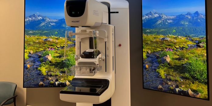
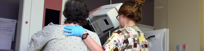
3D tomosynthesis Imaging – thinner, more defined images of breast
Annual screening mammograms
Diagnostic mammograms
The mammography suite was upgraded in 2024 through a generous donation by the
Horizon Health Foundation. Amenities include advanced lighting, customizable screens, and innovative elements designed
to enhance patient experience. The suite includes a private dressing room,
warm robes, and a recliner for added comfort.
We are one of the few critical access hospitals in Illinois that utilizes
artificial intelligence on all CT & MRI scans for higher quality images
and accuracy.
We share images with more than 62 hospitals & clinics across the Midwest,
reducing costs & enhancing patient care.
Our “low dose” testing reduces radiation exposure by as much as 60%.
Podcasts
Lung Scan
Emilee Campbell, radiology staff manager, explains the importance of a
lung scan and who should get one.
Coronary Calcium Scan
Emilee Campbell, radiology staff manager, explains the importance of a
coronary calcium scan – also known as a
heart scan – and who should get one.
Nuclear Stress Testing
Dr. Bruce Houle, full-time radiologist, discusses the importance of nuclear stress testing
and who can benefit from it.
Mammograms
Dr. Bruce Houle, full-time radiologist, discusses the importance of early screening for
breast cancer, screening recommendations, digital technology, and more.


.jpg)